
Health Library
Color vision test
Eye test - color; Vision test - color; Ishihara color vision test
A color vision test checks your ability to distinguish between different colors.
Images
I Would Like to Learn About:
How the Test is Performed
You will sit in a comfortable position in regular lighting. The health care provider will explain the test to you.
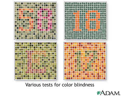
There are several tests for color vision. During the most common, you will be shown several cards with colored dot patterns. These cards are called Ishihara plates. In the patterns, some of the dots will appear to form numbers or symbols. You will be asked to identify the symbols, if possible.
As you cover one eye, the tester will hold the cards 14 inches (35 centimeters) from your face and ask you to quickly identify the symbol found in each color pattern.
Depending on the problem suspected, you may be asked to determine the intensity of a color, particularly in one eye compared to the other. This is often tested by using the cap of a red eyedrop bottle.
How to Prepare for the Test
If your child is having this test performed, it may be helpful to explain how the test will feel, and to practice or demonstrate on a doll. Your child will feel less anxious about the test if you explain what will happen and why.
Usually there is a sample card of multicolored dots that almost everyone can identify, even people with color vision problems.
If you or your child normally wears glasses, wear them during the test.
Small children may be asked to tell the difference between a red bottle cap and caps of a different color.
How the Test will Feel
The test is similar to a vision test.
Why the Test is Performed
This test is done to determine whether you have any problems with your color vision.
Color vision problems often fall into two categories:
- Present from birth (congenital) problems in the light-sensitive cells (cones) of the retina (the light-sensitive layer at the back of the eye) -- the color cards are used in this case.
- Diseases of the optic nerve (the nerve that carries visual information from the eye to the brain) -- the bottle caps are used in this case.
Normal Results
If your color vision is normal, you will be able to distinguish all colors.
What Abnormal Results Mean
This test can determine the following congenital (present from birth) color vision problems:
- Achromatopsia -- complete color blindness, seeing only shades of gray
- Deuteranopia -- difficulty telling the difference between red/purple and green/purple
- Protanopia -- difficulty telling the difference between blue/green and red/green
- Tritanopia -- difficulty telling the difference between yellow/green and blue/green
Problems in the optic nerve can show up as a loss of color intensity, although the color card test may be normal.
Risks
There are no risks with this test.
Related Information
Color blindnessReferences
Chuck RS, Dunn SP, Flaxel CJ; American Academy of Ophthalmology Preferred Practice Pattern Committee, et al. Comprehensive adult medical eye evaluation preferred practice pattern. Ophthalmology. 2021;128(1):1-29. www.aaojournal.org/article/S0161-6420(20)31026-5/fulltext. Published November 12, 2020. Accessed March 2, 2021.
Karepov S, Ellenbogen T. Metasurface-based contact lenses for color vision deficiency. Opt Lett. 2020;45(6):1379-1382. PMID: 32163975 pubmed.ncbi.nlm.nih.gov/32163975/.
Salmon JF. Hereditary fundus dystrophies. In: Salmon JF, ed. Kanski's Clinical Ophthalmology. 9th ed. Philadelphia, PA: Elsevier; 2020:chap 15.
Wallace DK, Morse CL, Melia M, et al; American Academy of Ophthalmology Preferred Practice Pattern Pediatric Ophthalmology/Strabismus Panel. Pediatric eye evaluations Preferred Practice Pattern: I. vision screening in the primary care and community setting; II. comprehensive ophthalmic examination. Ophthalmology. 2018;125(1):184-227. PMID: 29108745 pubmed.ncbi.nlm.nih.gov/29108745/.
BACK TO TOPReview Date: 3/2/2021
Reviewed By: Franklin W. Lusby, MD, Ophthalmologist, Lusby Vision Institute, La Jolla, CA. Also reviewed by David Zieve, MD, MHA, Medical Director, Brenda Conaway, Editorial Director, and the A.D.A.M. Editorial team.
 | A.D.A.M., Inc. is accredited by URAC, for Health Content Provider (www.urac.org). URAC's accreditation program is an independent audit to verify that A.D.A.M. follows rigorous standards of quality and accountability. A.D.A.M. is among the first to achieve this important distinction for online health information and services. Learn more about A.D.A.M.'s editorial policy, editorial process and privacy policy. A.D.A.M. is also a founding member of Hi-Ethics. This site complies with the HONcode standard for trustworthy health information: verify here. |
The information provided herein should not be used during any medical emergency or for the diagnosis or treatment of any medical condition. A licensed medical professional should be consulted for diagnosis and treatment of any and all medical conditions. Links to other sites are provided for information only -- they do not constitute endorsements of those other sites. © 1997- 2021 A.D.A.M., a business unit of Ebix, Inc. Any duplication or distribution of the information contained herein is strictly prohibited.
![]()